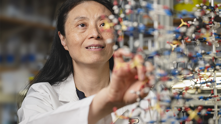
Purdue University researcher Danzhou Yang is exploring a natural check on cancer that could be used to stop the disease. (Purdue University photo/Charles Jischke)
Purdue researchers study natural curb on cancer as potential therapy
August 27, 2025 | Mary Martialay
WEST LAFAYETTE, Ind. — Could a natural check on cancer be used to stop the disease? It’s possible, but to leverage a system that nature designed, we must first understand it. Researchers led by a team at Purdue University are exploring a molecular mechanism that curbs the breakneck cell division associated with cancer. Their work opens the door to developing drugs that capitalize on the mechanism’s effects.
“We think this system evolved naturally in the context of cancer. It’s like a braking system. So now we’re asking, ‘How can we use this for drug discovery?’” said Danzhou Yang, the Martha and Fred Borch Chair in Cancer Therapeutics and Distinguished Professor of Medicinal Chemistry and Molecular Pharmacology in the College of Pharmacy’s Borch Department of Medicinal Chemistry and Molecular Pharmacology. The team’s most recent research appeared in the journal Science.
In images with resolution on the level of individual atoms, the researchers show how the brake works, with a protein binding to a segment of DNA associated with cancer like two Lego pieces snapped together. The DNA is part of the c-MYC gene, which, when improperly regulated, is the main driver of all blood cancers and 80% of human solid tumors. The MYC oncogene plays a significant role in cancer development and progression, acting as a “master regulator” that drives cell growth, proliferation and metabolism. While bound to the protein nucleolin, the c-MYC gene cannot be copied, blocking a necessary step in uncontrolled cell division.
Determining the structure of the complex that forms between the DNA and protein is an achievement on its own, but the unusual shape of the complex is equally enlightening. Yang’s team found that four modules on nucleolin, like beads on a string, bind to four connected loops of DNA in a globular structure that is entirely novel.
“The mechanism we’ve discovered is completely paradigm-changing. Nobody expected this binding mode of DNA,” Yang said. “Each globule binds to a loop weakly. But all together, you have this cooperative binding, which is very specific and very strong.”
Yang is a member of the Purdue Institute for Cancer Research and part of Purdue’s presidential One Health initiative, which involves research at the intersection of human, animal and plant health and well-being.
DNA encodes instructions for building proteins in a chain of only four molecules, called nucleotides, in a system similar to Morse code. When not in use, the two strands of DNA in the genome of a cell are zipped together, forming the familiar double helix. But throughout the life of the cell, the strands are constantly unzipped at intervals along their length, allowing the gene in one strand to be transcribed into RNA, the first step in expressing the gene to produce needed materials from the instructions in the DNA.
Some genes encode the instructions for materials that help the cell divide into two cells, a complex dance that is tightly controlled. Genetic mutations, environmental factors and changes that alter how often a particular gene is expressed can all disrupt that process, leading to out-of-control cell growth and division and cancer.
The c-MYC gene is part of that tight regulation. It encodes the instructions for producing the c-MYC protein, one of a family of proteins called transcription factors, which act like an on switch for many other genes by controlling the amount of protein expressed from that other gene. C-MYC helps to regulate cellular processes like cell growth, division and even immune response. Mutated forms of the c-MYC gene drive cancer by producing excessive amounts of the c-MYC transcription factor and disrupting the correct rhythm of transcription.
But somehow, said Yang, evolution has taken note and made a move to contain the problem. When unzipped, sections of DNA rich in the nucleotide guanine sometimes clump together, forming an odd temporary knot known as a G-quadruplex. Such regions are found in the c-MYC gene, and when the c-MYC gene are accessed at an abnormally rapid rate consistent with cancer, the G-quadruplex knot occurs with vastly greater frequency than in the healthy cells. But if nucleolin binds to the G-quadruplex, it holds the knot, blocking transcriptional machinery from binding to the gene and stopping transcription.
The reprieve is only temporary, as the G-quadruplex is short-lived. But it’s a working mechanism to stop cancer — and one that researchers can improve upon.
“If we can target this G-quadruplex, stabilize its complex with nucleolin, and make it much longer-lived, we can turn off c-MYC transcription. That can inhibit or even cure cancer,” Yang said.
C-MYC is well-known as a gene that contributes to the development of cancer, and Yang said it’s a hot target for drug discovery. The c-MYC G-quadruplex is a viable drug target, and knowing the exact three-dimensional structure of the complex between the G-quadruplex and nucleolin at the atomic level — how the Lego pieces snap together — makes it possible to design a drug that adheres to the G-quadruplex as well as nucleolin but lasts longer. The research published in Science presents that level of detail, as well as the surprising nature of the four-part binding between the modular protein and the loops of globular DNA.
Detailed structural illustrations show how the globular DNA in the G-quadruplex presents four loops that offer a docking site for proteins. Ordinarily, modular proteins like nucleolin form only a weak connection to DNA. But in this case, four modules on nucleolin bind to all four loops on the globular DNA.
“We can see how this modular protein binds to all four of the loops of the globular DNA G-quadruplex structures, creating a very strong binding,” Yang said. “And this is important not just to cancer, but to many other aspects of biology. The G-quadruplex can form in many guanine-rich sequences with significant functional roles.”
In further research, Yang will continue to gather details on how G-quadruplex interacts with other proteins to control transcription, what Yang calls “G-quadruplex-based epigenetic transcriptional control,” producing an even clearer picture for drug discovery. Armed with that information, her team will explore drugs that could bind to the G-quadruplex and associated proteins, searching for what Yang calls “inhibitors of oncogenic epigenetic transcriptional control.”
At Purdue, Yang was joined in the research by Nicholas Noinaj, an associate professor of biological sciences in the College of Science; current and former graduate students and postdoctoral fellows and experts from other institutions also contributed to this work.
The team’s article, “Structural basis for nucleolin recognition of MYC promoter G-quadruplex,” was produced with support from the National Institutes of Health, the Purdue Institute for Cancer Research and the Intramural Research Program of the National Cancer Institute.
About Purdue University
Purdue University is a public research university leading with excellence at scale. Ranked among top 10 public universities in the United States, Purdue discovers, disseminates and deploys knowledge with a quality and at a scale second to none. More than 107,000 students study at Purdue across multiple campuses, locations and modalities, including more than 58,000 at our main campus in West Lafayette and Indianapolis. Committed to affordability and accessibility, Purdue’s main campus has frozen tuition 14 years in a row. See how Purdue never stops in the persistent pursuit of the next giant leap — including its comprehensive urban expansion, the Mitch Daniels School of Business, Purdue Computes and the One Health initiative — at https://www.purdue.edu/president/strategic-initiatives.
Paper
Structural basis for nucleolin recognition of MYC promoter G-quadruplex
Science


